What a protrusion actually is
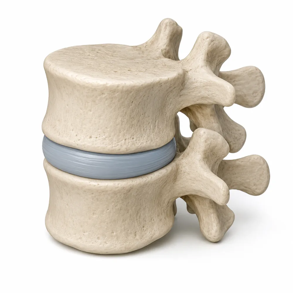
Your lumbar MRI report says disc protrusion at L4-L5, and the word alone is enough to make you sit differently for the rest of the day. It sounds like something broke.
The word describes a shape, not an injury and not a diagnosis.
Between each pair of vertebrae sits a disc: a tough outer ring with a softer centre, working as a cushion. When part of that disc pushes out past the edge of the vertebrae, radiologists describe how far it goes and what shape it makes.
The vocabulary is standardised. The reference is Lumbar Disc Nomenclature: Version 2.0, agreed between the North American Spine Society, the American Society of Spine Radiology and the American Society of Neuroradiology, and it defines three terms people constantly mix up:
- Bulge — the disc extends beyond the vertebral edge around more than 25% of its circumference, usually less than 3 mm out. It is generalised, not focal.
- Protrusion — a focal displacement, under 25% of the circumference, where the part sticking out is narrower than its base. Like a bud.
- Extrusion — the same thing but the part sticking out is wider than its base. Like a blob squeezed through a gap.
Protrusion and extrusion are both types of herniation. So "protrusion" already tells you it is the smaller-shaped of the two.
The number that puts it in perspective
This is the part almost nobody is told, and it changes how the report reads.
A systematic review published in the American Journal of Neuroradiology in 2015 pooled imaging from 3,110 people with no symptoms at all — no back pain, nothing. Among them:
- 29% of 30-year-olds had a disc protrusion. By age 80 it was 43%.
- Disc bulges went from 30% at age 20 to 84% at age 80.
- Disc degeneration went from 37% at age 20 to 96% at age 80.
Read that again: nearly a third of people in their thirties who feel perfectly fine have a disc protrusion sitting on their spine, undiscovered, causing nothing. The authors concluded that many of these findings are likely part of normal ageing and unassociated with pain.
That does not mean your protrusion is irrelevant. It means the image alone cannot tell you whether it is.
What decides whether it matters
Your doctor is comparing the picture with you. Three things matter more than the word itself:
- Does the location match your symptoms? A protrusion pressing a nerve root on the left explains left-leg pain. It does not explain right-leg pain.
- Is there contact with a nerve? The report usually says so: contacts the L5 nerve root, narrows the lateral recess, no neural compromise. That last one means it is not touching anything.
- What does the examination show? Strength, reflexes, sensation. That is what turns an image into a diagnosis.
This is exactly why so many of these reports end with "clinical correlation is recommended" — we wrote about that phrase here.
Questions worth taking to your appointment
- Does the level in the report match where my pain is?
- Does the report say anything about nerve contact or compression?
- Is this new, or was it in my previous scan too?
- Does this change what we do, or is it a finding we simply note?
That last one is the important one. A finding that changes nothing about your treatment is a finding you can stop worrying about.
Sources
- Brinjikji W. et al. Systematic Literature Review of Imaging Features of Spinal Degeneration in Asymptomatic Populations. American Journal of Neuroradiology, 2015. The figures for the 3,110 people without symptoms come from here.
- Fardon D.F. et al. Lumbar disc nomenclature: version 2.0. The Spine Journal, 2014. Joint recommendations of the North American Spine Society, the American Society of Spine Radiology and the American Society of Neuroradiology; this is what defines bulge, protrusion and extrusion.
This explains vocabulary, not your case. Nothing here can tell you whether your protrusion is causing your pain — that needs someone who can examine you and who knows your history.
What ExplainMyScan does is the step before that: you upload your report, say what worries you, and get it explained in plain language, so you walk into the appointment understanding what you are holding and knowing what to ask. It does not diagnose and it does not replace your doctor.
Frequently asked questions
- What does disc protrusion mean on an MRI report?
- It describes a shape, not an injury. Part of the intervertebral disc extends beyond the edge of the vertebrae in a focal way, under 25% of the disc circumference, and the part sticking out is narrower than its base.
- What is the difference between a bulge, a protrusion and a herniation?
- Under the Lumbar Disc Nomenclature 2.0, a bulge involves more than 25% of the circumference and extends a short distance. A protrusion is focal and narrower than its base. An extrusion is focal and wider than its base. Protrusion and extrusion are both types of herniation.
- Can you have a disc protrusion without pain?
- Yes, and it is common. A 2015 systematic review in the American Journal of Neuroradiology pooled imaging from 3,110 people with no symptoms and found disc protrusions in 29% of 30-year-olds, rising to 43% by age 80.
- Does a protrusion on my report explain my pain?
- The image alone cannot say. What matters is whether the level matches where your symptoms are, whether the report mentions nerve contact, and what a physical examination shows.